Diagram Of Upper Leg Muscles And Tendons : Lower Extremity Anatomy Bones Muscles Nerves Vessels Kenhub / Between the tendons is a space called the popliteal fossa, with a small fat pad.
Diagram Of Upper Leg Muscles And Tendons : Lower Extremity Anatomy Bones Muscles Nerves Vessels Kenhub / Between the tendons is a space called the popliteal fossa, with a small fat pad.. Foot muscles and tendons ã¢â?â? Many of the leg's muscles are also adapted to bipedalism, most substantially the gluteal muscles, the extensors of the knee joint, and the calf muscles.8. A muscle along the outside of the leg that bends the foot out at the ankle. Following injury ligaments and tendons may take a long time to heal because. Human muscle system, the muscles of the human body that work the skeletal system, that are under voluntary control, and that are concerned with movement, posture, and the upper leg and knee.
This study investigated the effects of upper extremity muscle fatigue on dynamic and static balance in young and old populations. Collectively, the muscles in this area in the lower part of the leg, the muscle belly combines with the soleus to from the calcaneal tendon, with inserts onto the calcaneus (the heel bone). Inferior surface of tarsals and metatarsals. Proximal phalanx of hallicus and tendon of extensor digitorum longus. Anatomy of leg and foot human muscular system.
Thigh Pain Injuries Symptoms Causes And Treatment from learn.ducklingdigital.co.uk In the lower leg, the anterior tibial enters the extensor compartment near the upper border of the interosseus membrane to descend between the. Other areas where tendonitis occurs include the hips and ankles. Between the tendons is a space called the popliteal fossa, with a small fat pad. This is where the gto comes into play. This muscle originates on the distal anterior surface of the fibula and the adjacent interossous membrane. The accompanying muscle diagram further reveals the positions of the muscles in this pose. Many of the leg's muscles are also adapted to bipedalism, most substantially the gluteal muscles, the extensors of the knee joint, and the calf muscles.8. Deep peroneal nerve supplies muscular branches to some parts of the leg and the ankle joint.
A tendon is the end part of a muscle that attaches the muscle to the bone.
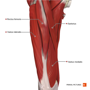
Created and produced by qa international. The muscle ends in tendons and the tendons plug the muscle into bones. Collectively, the muscles in this area in the lower part of the leg, the muscle belly combines with the soleus to from the calcaneal tendon, with inserts onto the calcaneus (the heel bone). This study investigated the effects of upper extremity muscle fatigue on dynamic and static balance in young and old populations. The accompanying muscle diagram further reveals the positions of the muscles in this pose. This is where the gto comes into play. Sartorius muscle appears from the anterior superior iliac spine and upper half of the notch immediately below it. Upper leg muscles and tendons. When studying the muscles of the leg, they can be compartmentalized into four primary groups: The anterior, lateral (fibular), superficial posterior, deep posterior compartments. Both peroneal tendons then course anteriorly toward the peroneal trochlea of the lateral. A tendinous intersection is usually observed about the middle of the muscle. Muscles in the arm diagram koibana info forearm anatomy upper limb anatomy arm muscle anatomy.
Following injury ligaments and tendons may take a long time to heal because. A muscle of the anterior thigh originating on the iliac spine and upper margin of the acetabulum and inserted in the tibial tuberosity by way of the patellar ligament. A muscle along the outside of the leg that bends the foot out at the ankle. Causes of upper leg pain related to trauma may include the following. When studying the muscles of the leg, they can be compartmentalized into four primary groups:
Inner Thigh Muscles Anatomy Anatomy Drawing Diagram from doctorlib.info When studying the muscles of the leg, they can be compartmentalized into four primary groups: Created and produced by qa international. The muscles of the foot mainly customize and improve the actions of the long tendons and help fine movements of the toes. Causes of upper leg pain related to trauma may include the following. The human body muscle anatomy body anatomy anatomy study muscular system bjorn borg human anatomy and physiology blood pressure remedies muscle building. The hamstring muscles are flexors, moving the upper leg (femur) at the hip joint and the lower leg (tibia and fibula). Upper limb trauma programme injuries. Section editor dean taylor, md.
When studying the muscles of the leg, they can be compartmentalized into four primary groups:
Not only are these groups located within the appropriate aspect of the tibia and fibula, but they are also defined by intermuscular. Back muscles diagram 12 photos of the back muscles diagram back muscle workout diagram, back muscles diagram for massage, back muscles diagram massage, human back muscles diagram, upper back muscles. Muscles in the arm diagram koibana info forearm anatomy upper limb anatomy arm muscle anatomy. Between the tendons is a space called the popliteal fossa, with a small fat pad. Tendonitis is usually seen after excessive repetitive movement with which the tendon gradually becomes tighter until the fibers start to tear. Lateral condyle and upper 2/3 of tibial shaft. When studying the muscles of the leg, they can be compartmentalized into four primary groups: This study investigated the effects of upper extremity muscle fatigue on dynamic and static balance in young and old populations. Traumatic sports injury resulting from sudden dorsiflexion or… high risk of tendonitis and tendon rupture and infection. Created and produced by qa international. Its tendon inserts on the dorsal surface of the base of 5th metatarsal bone. The accompanying muscle diagram further reveals the positions of the muscles in this pose. The muscles of the foot mainly customize and improve the actions of the long tendons and help fine movements of the toes.
Muscles in the arm diagram koibana info forearm anatomy upper limb anatomy arm muscle anatomy. Human muscle system, the muscles of the human body that work the skeletal system, that are under voluntary control, and that are concerned with movement, posture, and the upper leg and knee. The deep muscles that impact leg movement are generally smaller that those that are directly involved in flexing the knee. Tendonitis is usually seen after excessive repetitive movement with which the tendon gradually becomes tighter until the fibers start to tear. One more example is the large muscle group of the quadriceps, located on the front of the upper leg.
Quadriceps Muscle Strain Physiopedia from www.physio-pedia.com This muscle originates on the distal anterior surface of the fibula and the adjacent interossous membrane. This study investigated the effects of upper extremity muscle fatigue on dynamic and static balance in young and old populations. The hamstring muscles are flexors, moving the upper leg (femur) at the hip joint and the lower leg (tibia and fibula). Related online courses on physioplus. Causes of upper leg pain related to trauma may include the following. This is where the gto comes into play. The anterior, lateral (fibular), superficial posterior, deep posterior compartments. Some are small in length, and others are thinner and less bulky than muscles that extend or flex the knee or primary superficial veins of right thigh and leg.
The hamstring muscles are flexors, moving the upper leg (femur) at the hip joint and the lower leg (tibia and fibula).
Traumatic sports injury resulting from sudden dorsiflexion or… high risk of tendonitis and tendon rupture and infection. Sartorius muscle appears from the anterior superior iliac spine and upper half of the notch immediately below it. Each of these muscles is a discrete organ constructed of skeletal muscle tissue, blood vessels, tendons, and nerves. The anterior, lateral (fibular), superficial posterior, deep posterior compartments. Deep peroneal nerve supplies muscular branches to some parts of the leg and the ankle joint. Tendons arm wrist anatomy arm muscle anatomy anatomy and physiology. Its tendon inserts on the dorsal surface of the base of 5th metatarsal bone. Both peroneal tendons then course anteriorly toward the peroneal trochlea of the lateral. The biomechanical effects of stretching. The accompanying muscle diagram further reveals the positions of the muscles in this pose. This is where the gto comes into play. Created and produced by qa international. The upper part of the aponeurosis is curved backward over the upper edge of the tendon of the gracilis it is the great extensor muscle of the leg, forming a large fleshy mass which covers the front and sides of the femur.
The accompanying muscle diagram further reveals the positions of the muscles in this pose upper leg muscles and tendons. Foot muscles and tendons ã¢â?â?


0 Komentar